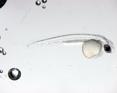
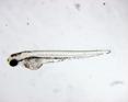
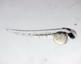
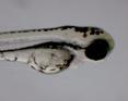
Zebrafish segmentation for high-throughput analysis
In today's biology, the zebrafish constitutes an ideal model for disease modeling and drug discovery since the zebrafish is vertebrate and its embryos develop quickly and outside the mother body. In this modeling, the data generated from large number of embryos become the bottleneck. To circumvent this issue, it is necessary to develop automated systems for zebrafish analysis. The first step of this analysis is to identify biological parts (head, tail, yolk sac, trunk, etc.) of a zebrafish. In this research project, you are expected to investigate image processing techniques for segmenting a zebrafish into its biological parts.
 |
 |
 |
 |
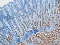
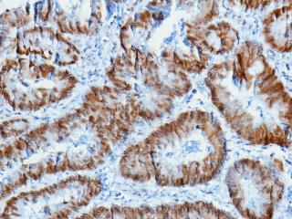
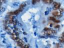
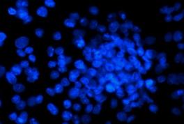
An automated system for immunohistochemistry
Immunohistochemistry is used to explore the molecular mechanisms on biopsies for identification and characterization of diseases including cancer. It relies on antibody-antigen reactions for identifying particular proteins in cells; the existence of a particular protein could be an indicator of a disease. The cells with positive reactions are stained brown whereas those without any reaction remain blue. Moreover, some antibodies lead to reactions in cell nuclei whereas others in cell cytoplasms. In order for a pathologist to make decisions, it is important to identify whether or not such reactions occur and if so, its intensity and whether it occurs in nuclei or cytoplasms. However, this process is subject to a considerable amount of observer variability. In this research project, you are expected to investigate different image processing techniques for implementing an automated analysis system. This system will focus on nuclei reactions, which requires identifying nuclear regions and quantifying the intensity of the reaction.
 |
 |
 |
 |
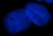
Segmentation and characterization of cells in fluorescence images for high-throughput screening
Automated fluorescence microscopy imaging is an important tool for drug discovery to obtain high-throughput screening. In this screening, it is essential to identify cells and compare the morphological changes in drug-treated cells against those of non-treated control cells. In this research project, you are expected to work on implementing an automated system for cell segmentation and characterization.
 |
 |
 |
 |